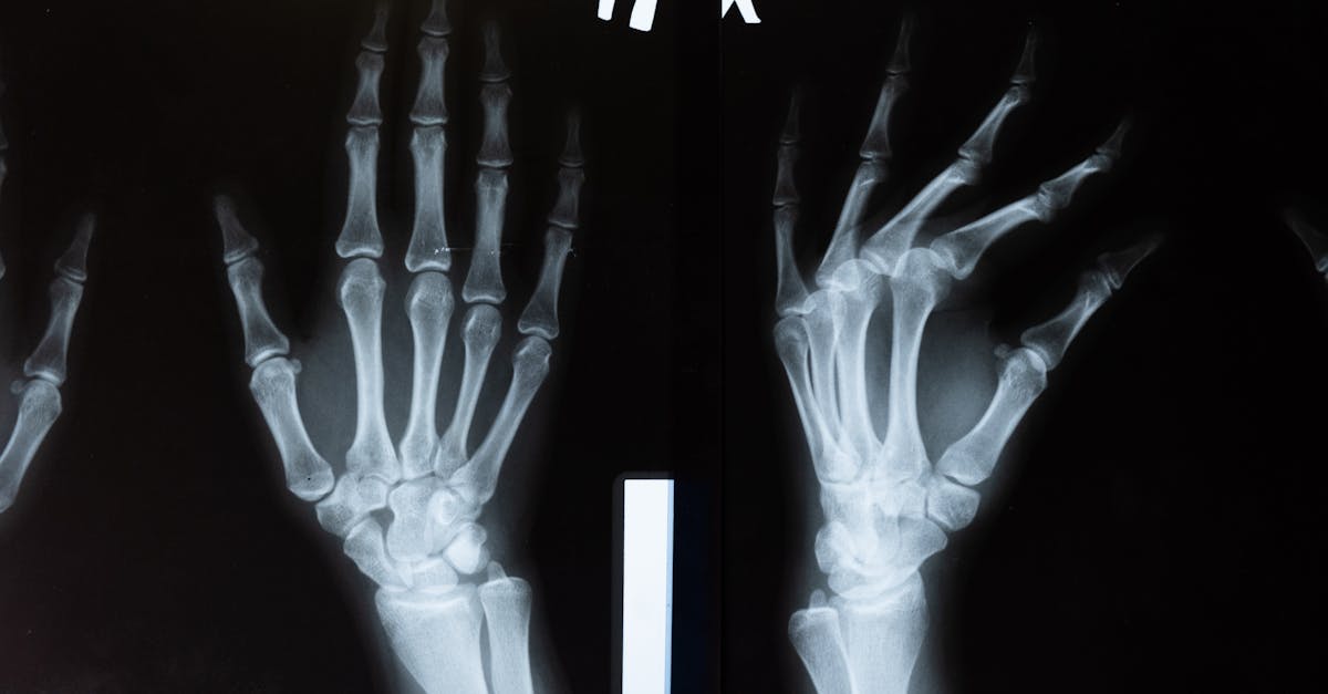
Medical imaging plays a crucial role in the diagnosis of heart diseases, facilitating the identification of various anomalies in the heart and blood vessels. Thanks to advanced techniques such as echocardiography, chest X-ray, and cardiac MRI, healthcare professionals can obtain precise images of the structure and cardiac function. These diagnostic tools not only allow for the detection of conditions such as heart failure and coronary artery disease but also enable the monitoring of existing conditions, thereby providing timely and appropriate care for affected patients.

Medical imaging is a fundamental pillar in the diagnosis and management of heart diseases. By allowing doctors to obtain precise visualization of cardiac structures and functions, it facilitates the diagnosis, assessment, and monitoring of cardiovascular pathologies. Understanding the different imaging techniques and their specific roles is essential for establishing an effective and tailored care pathway for each patient.
The techniques of cardiac imaging primarily include echocardiography, chest X-ray, cardiac CT, and cardiac MRI. Each of these methods provides distinct information that can together enable a comprehensive assessment of a patient’s cardiac health status. Due to their complementarity, the combined use of these techniques is often necessary for complete management.
Echocardiography is a non-invasive method that uses ultrasound to produce moving images of the heart. This technique allows for the assessment of cardiac structure, proper valve function, and blood flow. Thanks to advanced techniques like Doppler echocardiography, it is also possible to measure the speed of blood in the vessels and identify regional contraction anomalies, potential signs of coronary artery disease. Thus, echocardiography is particularly useful in diagnosing heart failure and in risk stratification in patients with cardiovascular symptoms.
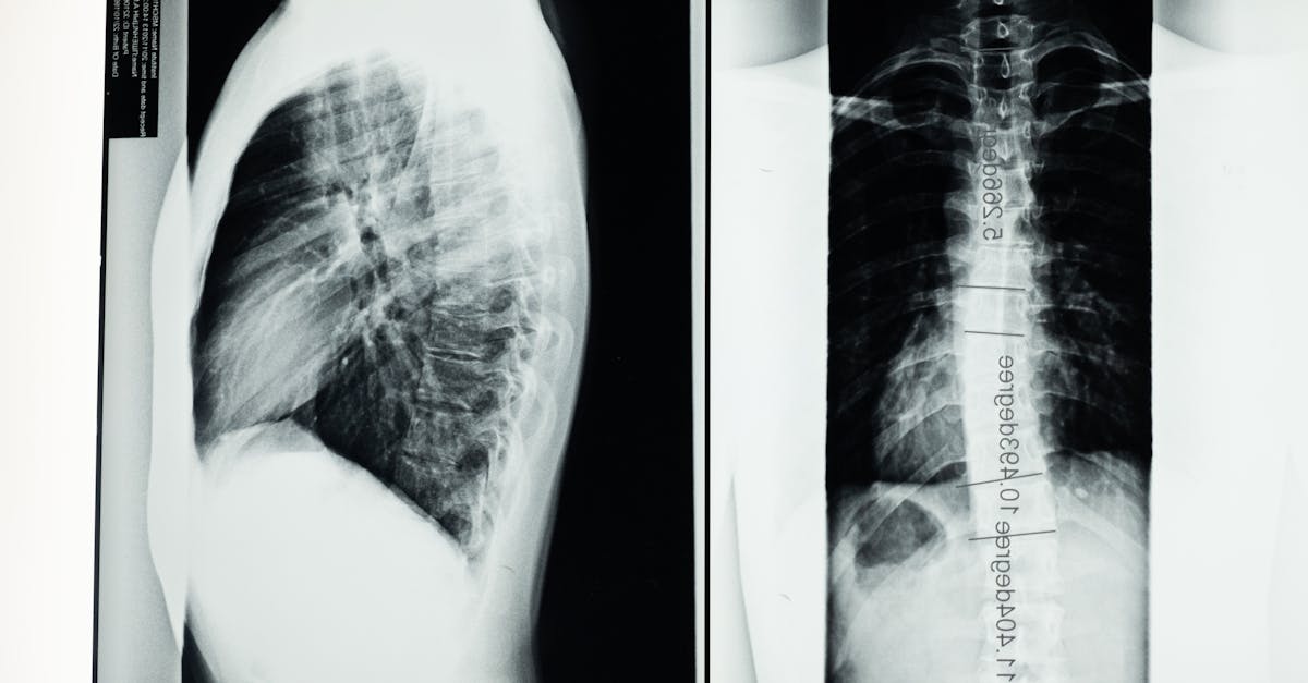
In addition to echocardiography, chest X-ray plays an important role in diagnosing cardiac disorders. Although it does not provide precise details on cardiac function, it can highlight anomalies such as cardiac dilatations or signs of acute heart failure. Furthermore, the X-ray can also help exclude other pulmonary pathologies that may involve the heart. Its use is therefore essential in a multimodal diagnostic approach.
The cardiac CT, or computed tomography, provides eclectic images of cardiac structures with excellent spatial resolution. This examination can detect coronary artery diseases and quantify the calcium score, a key indicator of the risk of cardiovascular events. Through this technique, it is possible to visualize the coronary arteries and identify anomalies such as stenoses, which may require protective intervention before serious complications occur.
Cardiac MRI, on the other hand, allows for high-quality images of cardiac structures, myocardial function, and blood flow. This technique is particularly valuable in detecting cardiomyopathies and congenital anomalies. By using it, clinicians can perform detailed evaluations of the left ventricular ejection fraction (LVEF), providing crucial data for risk stratification and therapeutic decision-making.
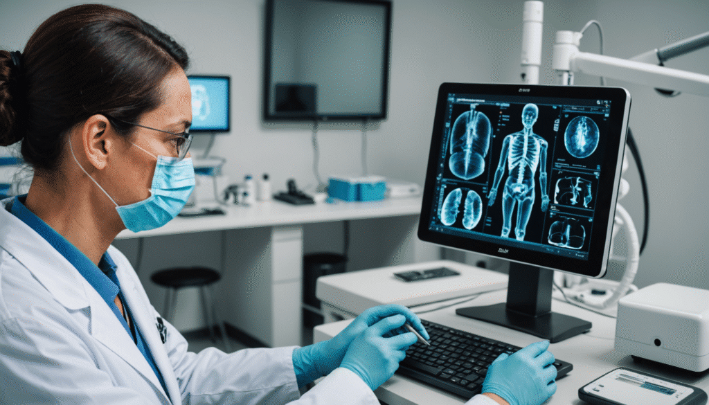
The integration of cardiac imaging into the care pathway is also driven by technological advancements, such as the emergence of artificial intelligence (AI) tools that support image interpretation. Studies have shown that these tools improve the accuracy and speed of diagnoses by enabling quantitative analysis of results and reducing the potential subjectivity associated with human interpretation. For instance, the implementation of AI in the evaluation of chest X-rays or cardiac ultrasounds transforms clinical practices and enhances their effectiveness.
These advancements in the field of medical imaging contribute to the democratization of access to cutting-edge technologies. It is essential that these techniques are available in various facilities, including in less urbanized areas, to ensure early diagnosis and prompt initiation of care. Moreover, the continuing education of healthcare professionals about these new technologies is inevitable. Understanding the operation and performance of these tools allows medical staff to utilize them optimally for the benefit of patients.
Finally, the potential of medical imaging is not limited solely to diagnosis. It also offers possibilities for treatment evaluation, disease monitoring, and improvement of clinical outcomes. Through precise visualization of therapeutic response, doctors can adjust treatments in real time, contributing to a better overall strategy for managing heart diseases. This includes, for example, the use of MRI to evaluate improvement in cardiac function after heart surgery or the optimization of drug treatment.
Ultimately, the role of medical imaging in the diagnosis of heart diseases is of paramount importance. By combining different imaging techniques, it is possible to make a complete and accurate diagnosis, assess the underlying mechanisms of diseases, and support patients throughout their care journey. Future developments in this field are promising and will continue to transform cardiology, strengthening the link between technology and clinical practice.
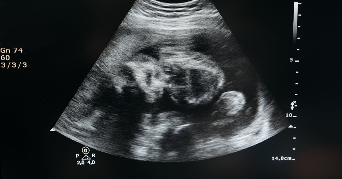
Medical imaging plays a fundamental role in the diagnosis of heart diseases. Through different techniques, it allows for precise visualizations of the structure and function of the heart, thus facilitating the identification of cardiovascular disorders and the development of appropriate treatment strategies. This text highlights the importance of these diagnostic techniques in the management of heart pathologies.
The main cardiac imaging techniques
The cardiac imaging examinations consist of several essential techniques. Among the most common are echocardiography, which uses ultrasound to visualize the heart in motion. This technique allows for the evaluation of the morphology of cardiac structures and their functionality. Another effective method is chest X-ray, which can assist in the diagnosis of heart failure by detecting changes in heart chambers and potential fluid accumulation in the lungs.
Cardiac MRI for advanced evaluation
Cardiac MRI is a leading non-invasive technique, particularly useful for analyzing cardiac tissues and assessing structural anomalies. It provides high-quality images, allowing for a better understanding of myocardial dysfunctions. MRI is also valuable for detecting pathologies such as cardiomyopathy, facilitating the measurement of myocardial fibrosis and the quantification of ventricular volume.
Use of Doppler ultrasound
Cardiac Doppler is also a key tool in medical imaging. Through this technique, it is possible to evaluate blood flow within the heart and vessels, thus identifying regional contraction anomalies that could indicate coronary artery disease. The results of the Doppler ultrasound are essential for risk stratification in patients presenting symptoms of heart failure.
The relevance of cardiac CT
The cardiac computed tomography (CT) is another effective diagnostic method, allowing examination of the coronary arteries and detection of potential arterial blockages. If coronary artery disease is suspected, this technique can provide an accurate evaluation of coronary risk. Additionally, CT can be used to measure the calcium score, which indicates the level of atherosclerosis in the arteries.
Integration of artificial intelligence
With technological advancements, the introduction of artificial intelligence (AI) in medical imaging is transforming the landscape of cardiac diagnosis. AI can quickly and accurately analyze medical images, thus improving the interpretation of results and shortening diagnostic time. This clinical decision support is particularly beneficial in managing complex heart diseases.
The various techniques of medical imaging prove to be indispensable for the diagnosis and management of heart diseases. They allow not only for the early detection of pathologies but also for effectively guiding appropriate medical interventions. Their integration with technological innovations also promises to further improve clinical outcomes in the future.

