THE new imaging-guided biopsy techniques represent significant advances in the field of diagnostic medicine. These methods minimally invasive improve the precision of tissue samples, allowing finer detection of pathologies while minimizing risks for the patient. Thanks to toolsadvanced imaging such as ultrasound and MRI, practitioners can now perform biopsies with guidance stereotaxic, thus offering a more targeted and effective approach. These innovations pave the way for early diagnosis, essential in the fight against diseases such as cancer.

Image-guided biopsy has been a major advance in the field of diagnostic procedures for several years. These enable tissue harvesting with increased precision, reducing the risks associated with more invasive surgical procedures. Thanks to cutting-edge technologies, these techniques offer valuable solutions in the early diagnosis of diseases such as cancer.
There guided biopsy is defined as a medical procedure allowing a tissue sample to be taken in order to submit it to histological analyses. The use of imaging is essential to minimize risk by enabling precise needle positioning. The imaging modalities used may vary, includingultrasound, there CT scan (scan) and theMRI (magnetic resonance imaging).
THE ultrasound-guided biopsies have gained popularity, particularly due to their non-invasiveness. This technique uses sound waves to locate an abnormality, thereby guiding the needle. It is particularly effective for breast and prostate biopsies. During an ultrasound-guided breast biopsy, the doctor inserts a fine needle under ultrasound guidance to reach the suspicious tissue. Thanks to this technique, samples can be taken with a very low complication rate.
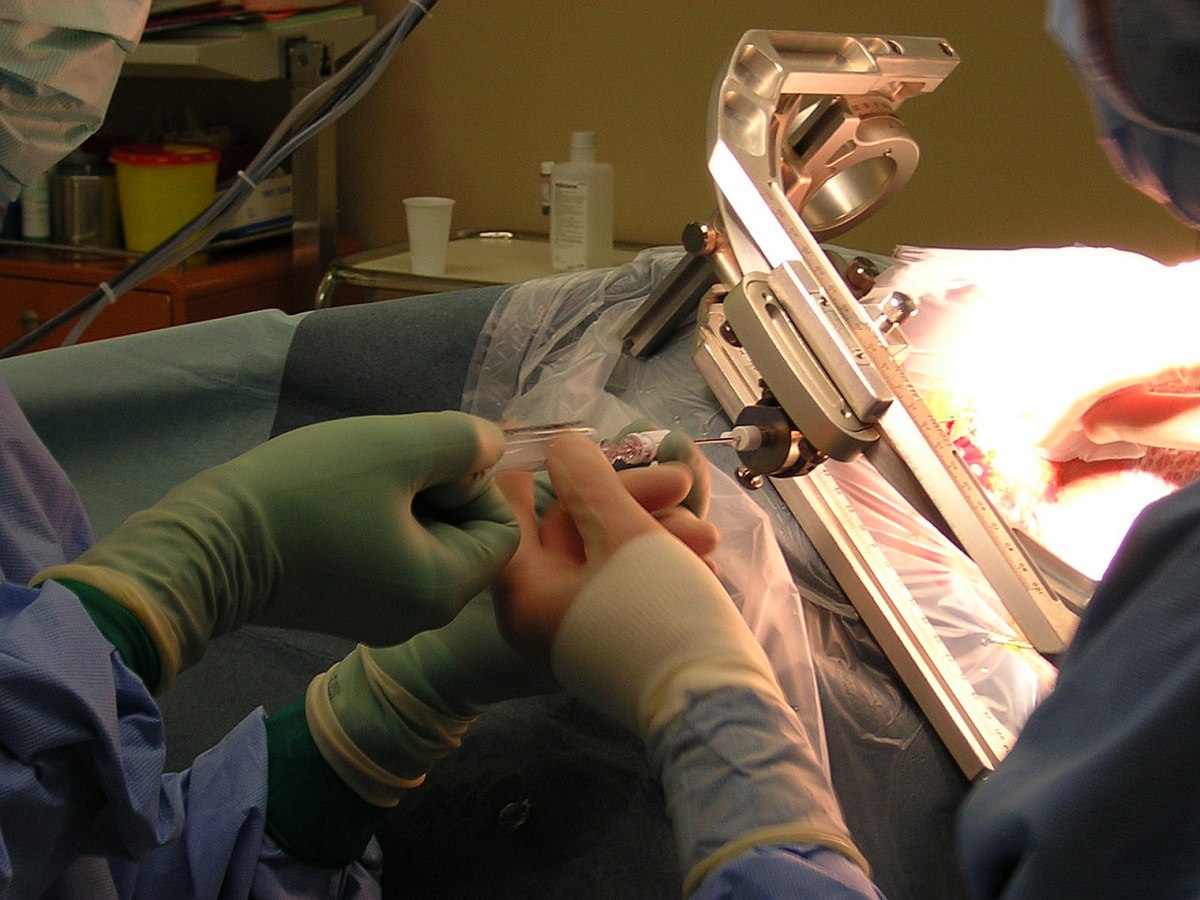
At the same time, the core biopsy under stereotaxic guidance also saw a significant improvement. This method relies on three-dimensional imaging systems that provide precise mapping of the area to be biopsy. Using imaging techniques such as ultrasound or CT scanning, the doctor can detect abnormalities that are difficult to see with traditional methods. Stereotactic drilling makes it possible to reach areas that would otherwise be inaccessible, while ensuring a sufficiently large sample for correct analysis.
The techniques of image merger represent another notable advance in the field of biopsies. By combining MRI and ultrasound data, it is possible to significantly improve tumor detection, particularly in the case of prostate cancer. This approach allows for a more precise visualization of the lesions, which increases the chances of successful sampling. There MRI-ultrasound fusion guided biopsy is increasingly common in clinical practice, thus improving patient care.
A project funded by the European Union recently contributed to the implementation of an innovative breast biopsy system guided by MEP imaging (preclinical evaluation model). This system uses advanced imaging techniques to detect breast abnormalities, enhancing the efficiency of biopsies while minimizing discomfort for patients. Thanks to this innovation, results can be obtained more quickly, thus providing a direct benefit to patients with suspicious lesions.
THE CT-guided biopsies for lung injuries also illustrate the progress made in this area. By using CT scanning to guide the sample, doctors can target lung nodules with unparalleled precision. This technique has been essential for the early diagnosis of lung cancers, allowing the disease to be detected at stages where treatment can be most effective.
There liquid biopsy is another method that has emerged as a promising alternative to traditional tissue biopsy. Although not directly an image-guided biopsy technique, it is worth mentioning because of its disruptive potential. Analyzing blood samples can detect circulating tumor cells and obtain information about tumor biology without the need for invasive intervention. This could represent an important advance in the monitoring of cancer patients.
The evolution of good practices for image-guided prostate biopsy has also been marked by an increasing integration of anatomical and functional information from imaging modalities. By combining T2-weighted images from MRI with information from isolated biopsies, doctors achieve an increasingly high level of precision and safety when taking samples.
Finally, the latest endoscopic techniques for the diagnosis of digestive diseases complete this panorama. Procedures such as colonoscopy Image-assisted imaging helps identify lesions within the gastrointestinal tract and perform targeted biopsies during the exam, reducing the number of separate procedures required.
New imaging-guided biopsy techniques are fundamentally changing the way diagnosis is made, making procedures safer and more reliable. This trend toward minimally invasiveness, combined with the use of advanced imaging technologies, offers new hopes for early diagnosis and intervention in the fight against various forms of disease.
For an overview of the different types of biopsies and their indications, it is possible to consult the resources available through this link: The different types of biopsies and their indications. Additionally, to learn more about advances in endoscopic techniques, visit: The latest endoscopic techniques for the diagnosis of digestive diseases.

Image-guided biopsy techniques represent a major advance in the field of medicine. These minimally invasive methods allow tissue collection with a high degree of precision while minimizing risk to the patient. Thanks to the use of sophisticated imaging tools, such as ultrasound, MRI and image fusion technologies, these new practices are continually improving and offer new perspectives for the diagnosis of various pathologies, especially cancers. This article explores the main imaging-guided biopsy techniques and their specific clinical applications.
Ultrasound-guided biopsy
Ultrasound-guided biopsy is a widely used method for collecting tissue samples from the breast and other organs. It uses ultrasound to visualize the target abnormality in real time, allowing precise guidance of the sampling needle. This technique is particularly appreciated for its speed and effectiveness, resulting in less pain and complications compared to more invasive methods, such as open surgery. By integrating standardized protocols, doctors can improve results and reduce false negatives during sampling.
Stereotactic breast biopsy
Stereotactic breast biopsy is an approach that uses mammographic images to locate and collect tissue samples. This technique is essential for detecting non-palpable abnormalities in breast tissue. By combining mammographic images with a three-dimensional coordinate system, it provides precise and targeted results, reducing the need for more invasive surgical procedures. Advances in the field of imaging technologies have made it possible to optimize this technique, making it more accessible.
MRI-guided biopsy
MRI-guided biopsy constitutes a cutting-edge technique for the diagnosis of cancers, particularly prostate cancer. It uses detailed images obtained by magnetic resonance to locate lesions. The superior image quality allows doctors to precisely target suspicious areas. Recently, the integration of MRI-ultrasound image fusion has significantly improved the detection and diagnosis of abnormalities, by combining the functional information of MRI with the visual reality of ultrasound. This approach has proven effective in improving the accuracy of prostate biopsies.
Core biopsy under stereotaxic guidance
This technique involves the use of a stabilized drilling system that allows the practitioner to collect tissue samples from hard-to-reach regions. Thanks to three-dimensional images, it guarantees a targeted approach and minimizes trauma to surrounding tissues. This method is particularly useful in cases where other biopsy techniques might not be feasible or might carry increased risks of complications.
The advantages of new guided biopsy techniques
Technological advances in image-guided biopsy bring many benefits, including better cancer detection and a reduction in invasive surgical procedures. For example, liquid biopsy, an emerging technique that analyzes blood samples for the presence of tumor cells, offers a non-invasive and less stressful approach for patients. This could revolutionize the early diagnosis of cancers.
New imaging-guided biopsy techniques are changing the landscape of medical diagnosis. By using advanced visualization methods, they enable more precise samples, thus contributing to earlier detection and monitoring of cancers. With the continued prospect of innovation, the future of guided biopsy looks bright, with solutions that could further improve patient care.


